Hepatic Hemangioma
Posted in General Ultrasound on June 16, 2014 by m.khodeer
RUQ US: 35 year old female, vague abdominal pain
Findings?
DDx?
further recommended studies, if any?
Additional Imaging (different patient)
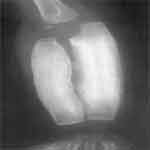
Contrasted CT, 120 s Delay
Same study, 300 s Delay
Final Dx?
Answers
Hepatic Hemangiomas


Ultrasound shows multiple homogeneous, hyperechoic hepatic masses: DDx hemangioma vs. more malignant process
Dynamic CT scanning (in a different patient) shows a well-circumscribed, hypoattenuating lesion that completely fills-in at 5 min
Answers
Hepatic Hemangioma – MRI Correlation
T2-weighted MRI:
Well-defined lesion, brighter than spleen
T1-Gad, at 30 s:
Nodular enhancement at periphery
T1 –Gad, AT 5 MIN:
Most of lesion has enhanced


Appearance of hemangiomas on US is characteristic but not pathognomonic
DDx includes Hemangioma, Mets, HCC, and nodular fat
Some feel patients at low risk for malignancy require no f/u, but at the least, 6-mo f/u US to evaluate stability is prudent. Nucs / CT /MRI can confirm. Most common benign livertumor, rarely symptomatic, typically no flow by US
Kasabach-Merritt syndrome: liver hemangiomas + platelet sequestration
Biopsy OK if there is normal liver between lesion and liver capsule
 Teaching Files


