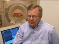
According to Raphael Bueno, MD, of Brigham and Women's Hospital in Boston, a new method that brings CT imaging into the operating room will enable surgeons to specifically isolate and remove small sub-centimeter lung nodules, leaving as much healthy tissue as possible.
His team presented their findings of this late-breaking research at the 94th AATS Annual Meeting in Toronto, ON, Canada on April 30, 2014.
To this day, lung cancer remains the deadliest cancer and a recent study, the National Lung Cancer Screening Trial, specifies that screening with low-dose computed tomography (CT) scans in smokers, who have certain risk factors, may decrease the number of deaths.
Lung cancer screening with CT can make out numerous small lung lesions that could possibly turn cancerous and should be removed surgically. The overall objective is to remove these small lung cancers but at the same time spare as much healthy lung as much as possible. In order to do so requires being able to accurately determine the precise location of the nodule and its margins.
"These results are exciting and promising, indicating that image-guided lung surgery could play a significant role in the treatment of lung cancer. This surgical approach has the potential to increase accuracy and reduce errors. It is like using GPS to navigate to the destination and perform a true surgical strike," said Bueno.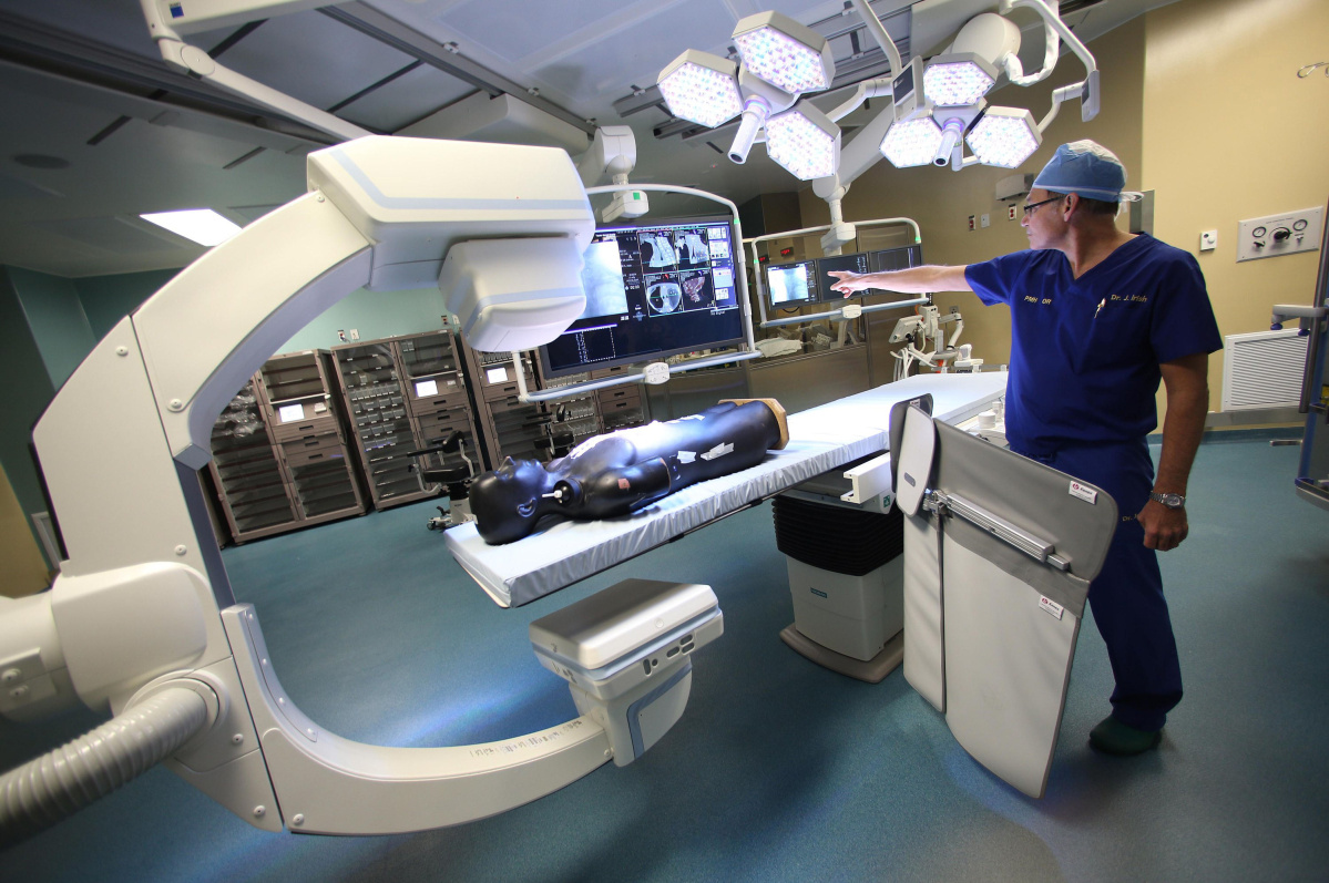
In this phase I/II clinical study conducted in collaboration with researchers from the Siemens Corporation, 20 patients were registered who had small pulmonary nodules in the outer half of the lung. Prior CT scans revealed that the lesions were very small, ranging from 0.6 to 1.8 cm. The nodules were so small that they could not be easily palpated or seen.
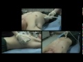
By employing a CT scanner in the operating room, surgeons first marked the location of the lung nodules by inserting two small markers (T-bars) through the skin and placing them next to the nodule. The markers have attached wires that make them visible to surgeons during the resection process. This technique is safe and successful for nodule localization and all patients underwent complete removal of the lesions with minimal removal of healthy lung tissue.
"We propose that image-guided video assisted thoracic surgery (IVATS) can be used to improve the ability to precisely identify small pulmonary nodules and allow for resections of sub-centimeter nodules," said Bueno.