


Researchers from the Georgia Institute of Technology have creating the technology for a catheter-based device that would provide insightful, real-time, three-dimensional (3D) imaging from inside the heart, coronary arteries, and peripheral blood vessels. With its all encompassing imaging, the novel device could better aid surgeons when working in the heart, and possibly allow more of patients' clogged arteries to be cleared without major surgery.
The device incorporates ultrasound transducers with processing electronics on a single 1.4 millimeter silicon chip. On-chip processing of signals permits data from more than a hundred elements on the device to be transmitted using just 13 tiny cables, enabling it to easily travel through circuitous blood vessels. The insightful images produced by the device would offer extensively more information than current cross-sectional ultrasound could or can.
Researchers have developed and tested a model able to present image data at 60 frames per second, and plan next to perform animal studies that could result in commercialization of the device.
"Our device will allow doctors to see the whole volume that is in front of them within a blood vessel. This will give cardiologists the equivalent of a flashlight so they can see blockages ahead of them in occluded arteries. It has the potential for reducing the amount of surgery that must be done to clear these vessels,” said, professor in the George W. Woodruff School of Mechanical Engineering at the Georgia Institute of Technology, F. Levent Degertekin.
Findings and details of the research were published online in the February 2014 issue of the journal IEEE Transactions on Ultrasonics, Ferroelectrics and Frequency Control.
Research leading to the development of the device was supported and funded by the National Institute of Biomedical Imaging and Bioengineering (NIBIB), part of the National Institutes of Health.
"If you're a doctor, you want to see what is going on inside the arteries and inside the heart, but most of the devices being used for this today provide only cross-sectional images. If you have an artery that is totally blocked, for example, you need a system that tells you what's in front of you. You need to see the front, back and sidewalls altogether. That kind of information is basically not available at this time,” said Degertekin.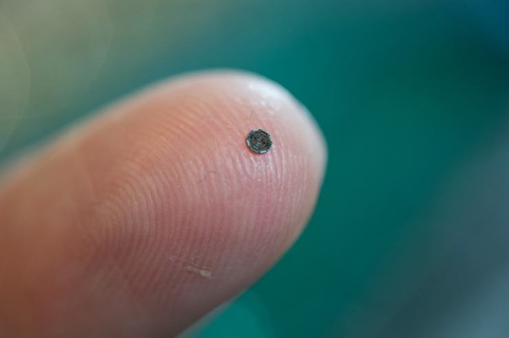
The single chip device integrates capacitive micromachined ultrasonic transducer (CMUT) arrays with front-end CMOS electronics technology to provide 3D intravascular ultrasound (IVUS) and intracardiac echography (ICE) images. The dual-ring array includes 56 ultrasound transmit elements and 48 receive elements. When gathered, the donut-shaped array is just 1.5 millimeters in diameter, with a 430-micron center hole to house a guide wire.
Power-saving circuitry in the array shuts down sensors when they are no longer required, enabling the device to function with just 20 milliwatts of power, decreasing the amount of heat generated inside the body. The ultrasound transducers operate at a frequency of 20 megahertz (MHz).
Imaging devices working from within blood vessels can offer higher resolution images than devices working from outside the body because they can function at higher frequencies. However, operating inside blood vessels calls for devices that are tiny and flexible enough to travel through the circulatory system. They must also be able to operate in blood.
Performing such a task requires a large number of factors to transmit and receive the ultrasound information. Transmitting data from these factors to external processing equipment could involve many cable connections, potentially limiting the device's ability to be threaded inside the body.
Degertekin and his collaborators have taken not and have addressed such a challenge by miniaturizing the elements and carrying out some of the processing on the probe itself, allowing them to attain what they believe are clinically-useful images with only 13 cables.
"You want the most compact and flexible catheter possible. We could not do that without integrating the electronics and the imaging array on the same chip,” Degertekin explained.
Based on their mock-model, the researchers anticipate to perform animal trials to showcase the device's potential applications. They ultimately expect to license the technology to an established medical diagnostic firm to conduct the clinical trials necessary to obtain FDA approval.
In the meantime, Degertekin hopes to develop a version of the device that could guide interventions in the heart under magnetic resonance imaging (MRI). Other plans include further reducing the size of the device to place it on a 400-micron diameter guide wire.


